


Nathalie Limodin
Home
Microstructure - fatigue damage mechanisms relationship: coupling X-ray tomography and field measurements
X-ray tomography allows me to follow in-situ microstructural evolutions and damage mechanisms within metallic materials and to revisit long-standing issues.
For example, synchrotron tomography has allowed us to analyze microstructural evolutions occurring during the solidification of "model" Al-Cu alloys and to identify the mechanisms acting at the scale of Al dendrite arms to test the validity of existing models [1-3].
Tomography also makes it possible to characterize the microstructure of an automotive industrial alloy (AlSi7Cu3) and to study the process-microstructure relationship.
The lost foam casting process, which is used for the manufacture of cylinder heads, induces pores and micro-shrinkages up to 1mm in size. The use of pore size distributions obtained by tomography has improved the relevance of lifetime probabilities calculated by an energy fatigue criterion [4]. The interdendritic space then contains three rigid phases: Cu-Fe intermetallics and eutectic Si. Intermetallic networks can be characterized in laboratory tomography, which makes it possible to quantify the influence of heat treatment or composition variation on the microstructure [5].
An understanding of the damage mechanisms is impossible without taking into account the complexity of the 3D microstructure. Indeed, in the presence of foundry defects, a crack can initiate internally w most observations are restricted to the surface. Thus, understanding the damage scenario requires coupling in-situ observations on the scale of the 3D microstructure with field measurements.
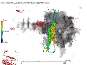
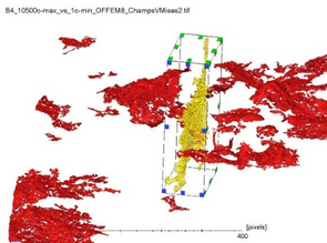
When the 3D microstructure (graphite nodules in spheroidal graphite cast iron, rigid phases and pores in an aluminum alloy) can be used as a speckle pattern for digital volume correlation (DVC), in-situ tomography tests not only allow identifying mechanisms but also quantifying them. The 3D displacement fields obtained by DVC during a 3D crack growth test in spheroidal graphite cast iron allowed us to extract the values of the stress intensity factor along the crack front and to study in particular the influence of the crack geometry on the opening and propagation [6]. In the AlSi7Cu3 alloy, the speckle pattern formed by the microstructure provides access to the deformation fields in the vicinity of the pores and a good agreement is observed between a finite element calculation performed on a 3D numerical model of the pores in the aluminium matrix and the field measurement during a monotonous tensile test under laboratory tomography [7]. Crack initiation occurs in the vicinity of pores where the measured deformations are the highest. To achieve scanning times compatible with low cycle fatigue tests, in-situ tests were performed under synchrotron tomography at room temperature but also at 250°C [8]. A high spatial resolution of the DVC can be obtained at a level of uncertainty that remains low [9], which makes it possible to measure deformation within the different phases. The cracks observed generally start internally near areas of high pore curvature [7]. Then they propagate according to the rigid inclusions where the cumulative von Mises deformation increases until rupture. This scenario depends on temperature.
N. Limodin, L. Salvo, M. Suéry, M. DiMichiel, Acta Mater. 55 (2007) 3177–3191.- N. Limodin, L. Salvo, E. Boller, M. Suéry, M. Felberbaum, S. Gailliègue, K. Madi, Acta Mater. 57 (2009) 2300–2310.
- L. Salvo, M. Suéry, A. Marmottant, N. Limodin, D. Bernard, Comptes Rendus Phys. 11 (2010) 641–649.
- F. Szmytka, N. Limodin, L. Wang, P. Osmond, J. Adrien, E. Charkaluk, J.Y. Buffiere, in:, J.Y. Buffière, M. Brune, F. Morel, Y. Nadot (Eds.), FDMD II - JIP 2014 - Fatigue Des. Mater. Defects, MATEC Web of Conferences, Paris, 2014, p. 05005.
- Z. Li, Influence of the Microstructure on Mechanical Properties and Damage Mechanisms in Al-Si-Cu Alloys by Using 2D and 3D in-Situ Analysis, Lille 1, 2016.
- N. Limodin, J. Réthoré, J.-Y. Buffière, F. Hild, S. Roux, W. Ludwig, J. Rannou, A. Gravouil, Acta Mater. 58 (2010) 2957–2967.
- L. Wang, N. Limodin, A. El Bartali, J.-F. Witz, R. Seghir, J.-Y. Buffiere, E. Charkaluk, Mater. Sci. Eng. A 673 (2016) 362–372.
- S. Dezecot, J.-Y. Buffiere, A. Koster, V. Maurel, F. Szmytka, E. Charkaluk, N. Dahdah, A. El Bartali, N. Limodin, J.-F. Witz, Scr. Mater. 113 (2016) 254–258.
- N. Dahdah, N. Limodin, A. El Bartali, J.F. Witz, R. Seghir, E. Charkaluk, J.Y. Buffiere, Strain 52 (2016) 324–335.